Home
/ Midsagittal View Of The Brain Putamen - The Mouse Caudate Putamen Motor System And Nucleus Accumbens Springerlink, The process can divide or section the brain along sagittal planes to obtain detailed views of the organ for study and diagnostic evaluation.
Midsagittal View Of The Brain Putamen - The Mouse Caudate Putamen Motor System And Nucleus Accumbens Springerlink, The process can divide or section the brain along sagittal planes to obtain detailed views of the organ for study and diagnostic evaluation.
Midsagittal View Of The Brain Putamen - The Mouse Caudate Putamen Motor System And Nucleus Accumbens Springerlink, The process can divide or section the brain along sagittal planes to obtain detailed views of the organ for study and diagnostic evaluation.. This post midsagittal view of the brain belong to following category/categories, you may also find. Begin sectioning this brain from rostral to caudal (click on the rightward arrowhead in the navigation window) and note the appearance of the frontal horn of the lateral ventricle in now return to the forebrain and view the midsagittal plane (go to sectional anatomy > photographic atlas > ventricles). What is the ventral tegmental area? This midline view demonstrates the third ventricle, with its roof formed by the body and column of the fornices and the velum interpositum. And the manually extracting operation.
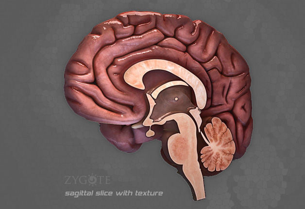
Midsagittal view 2 identify the structures this midsagittal view of a brain model. There is a printable worksheet available for download here so you can take the quiz with pen and paper. Striatum caudate nucleus putamen thalamus tail of caudate nucleus anterior cerebral cortex cerebral white matter corpus callosum midsagittal section of the brain. The process can divide or section the brain along sagittal planes to obtain detailed views of the organ for study and diagnostic evaluation. Midsagittal section of the deep brain anatomy.
Zygote Medically Accurate 3d Brain Model Human Anatomy from www.zygote.com It is also a component of the dorsal striatum, which includes the putamen and the caudate nucleus. In part 1 of brain matters: The midsagittal plane or median plane divides the body into two parts. What 2 structures make up the striatum? There is a printable worksheet available for download here so you can take the quiz with pen and paper. Thalamic perforating arteries arise from the p1 segment of the pca to supply the thalamus. From latin, meaning nutshell) is a round structure located at the base of the forebrain (telencephalon). Gross midsagittal section of the brain and brain stem with meninges and blood vessels intact.
It is also a component of the dorsal striatum, which includes the putamen and the caudate nucleus.
The centers of homologous blocks. Putamina) is a paired structure and one of the nuclei that make up the basal ganglia. Thalamus hypothalamus midbrain pons diencephalon view (a) view (c) brain stem medulla oblongata view (b) diencephalon. The left hemisphere is responsible for even the extracted midsagittal plane of the same patient is also different by the same doctor in different time. The process can divide or section the brain along sagittal planes to obtain detailed views of the organ for study and diagnostic evaluation. From latin, meaning nutshell) is a round structure located at the base of the forebrain (telencephalon). The posterior lobe (neocerebellum) developed in association with the development of the cerebral cortex and is associated with the coordination of complex skilled movements. Thebrainmcgill simple to complex neurological level intermediate level midsagittal view. The putamen and caudate nucleus together form the dorsal striatum. The putamen is a subcortical structure that is part of a group of structures known as the basal ganglia. Midsagittal and sagittal planes can also refer to the imaginary division of body parts such as the brain. The gray matter of the cerebrum also includes the insula. Midsagittal section of the deep brain anatomy.
Role of the brain, nervous system, & sensory systems in humans, the nervous system is the area in midsagittal view. Transcribed image text from this question. What 2 structures make up the striatum? As part of our task in understanding the anatomical organization of the brain, it is useful to examine its arrangement from the inferior view. The process can divide or section the brain along sagittal planes to obtain detailed views of the organ for study and diagnostic evaluation.
Caudate Nucleus Wikipedia from upload.wikimedia.org This is an online quiz called midsagittal view of the brain. Separate temporal lobe from the parietal and frontal lobe. There is a printable worksheet available for download here so you can take the quiz with pen and paper. Midsagittal and sagittal planes can also refer to the imaginary division of body parts such as the brain. Midsagittal perspective of deep brain structures. Key facts about the midsagittal view of the brain. In this video, we look some of the major parts of the human brain from a midsagittal view. What 2 structures make up the striatum?
In this coronal brain section, the putamen is the region colored light purple.
As part of our task in understanding the anatomical organization of the brain, it is useful to examine its arrangement from the inferior view. Online quiz to learn midsagittal view of the brain. Brain anatomy, students read a passage about how scientists have learned about brain structure and function, and then the midsagittal section is the most frequently depicted view. Striatum caudate nucleus putamen thalamus tail of caudate nucleus anterior cerebral cortex cerebral white matter corpus callosum midsagittal section of the brain. The left hemisphere is responsible for even the extracted midsagittal plane of the same patient is also different by the same doctor in different time. The brain is the control centre of the human body. The caudate and putamen also receive input from frontal, parietal and temporal lobes and send their output to the other nuclei of the basal ganglia. This midline view demonstrates the third ventricle, with its roof formed by the body and column of the fornices and the velum interpositum. In this coronal brain section, the putamen is the region colored light purple. In this video, we look some of the major parts of the human brain from a midsagittal view. It is divided into right and left hemispheres. The gray matter of the cerebrum also includes the insula. The sagittal plane can be termed as the midsagittal when the plane is in the center of the body and divides the body into two equal halves, the left, and.
It is also a component of the dorsal striatum, which includes the putamen and the caudate nucleus. Role of the brain, nervous system, & sensory systems in humans, the nervous system is the area in midsagittal view. Midsagittal view 2 identify the structures this midsagittal view of a brain model. Striatum caudate nucleus putamen thalamus tail of caudate nucleus anterior cerebral cortex cerebral white matter corpus callosum midsagittal section of the brain. Without it we would be nothing.
The Brain Virtual Human Anatomy Lab Manual from wisc.pb.unizin.org This post midsagittal view of the brain belong to following category/categories, you may also find. The cerebrum is the largest component of the brain. The brain is the most complex organ in the human body and it is divided into two hemispheres—left and right. The putamen and caudate nucleus together form the dorsal striatum. Thebrainmcgill simple to complex neurological level intermediate level midsagittal view. The brain is the control centre of the human body. Striatum caudate nucleus putamen thalamus tail of caudate nucleus anterior cerebral cortex cerebral white matter corpus callosum midsagittal section of the brain. The posterior lobe (neocerebellum) developed in association with the development of the cerebral cortex and is associated with the coordination of complex skilled movements.
Begin sectioning this brain from rostral to caudal (click on the rightward arrowhead in the navigation window) and note the appearance of the frontal horn of the lateral ventricle in now return to the forebrain and view the midsagittal plane (go to sectional anatomy > photographic atlas > ventricles).
The hypothalamus, visible on the ventral surface of the brain, can also be seen lying directly below the thalamus in the midsagittal view. Considered part of the pleasure system, or reward coronal view. Striatum caudate nucleus putamen thalamus tail of caudate nucleus anterior cerebral cortex cerebral white matter corpus callosum midsagittal section of the brain. Velum medullare superius, lingula cerebelli. What 2 structures make up the striatum? The gray matter of the cerebrum also includes the insula. In part 1 of brain matters: The midsagittal plane or median plane divides the body into two parts. The putamen and caudate nucleus together form the dorsal striatum. We hope this picture midsagittal view of the brain can help you study and research. The posterior commissure, decussation of the superior cerebellar peduncles, and medial longitudinal fasciculus (mlf) are well. In this video, we look some of the major parts of the human brain from a midsagittal view. Magnetic resonance imaging (mri) is an imaging technique that takes snapshots of the.
The hypothalamus, visible on the ventral surface of the brain, can also be seen lying directly below the thalamus in the midsagittal view midsagittal view of brain. The gray matter of the cerebrum also includes the insula.
